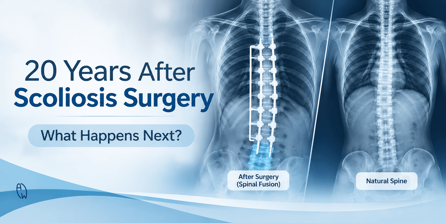
What Happens to Your Spine 10–20 Years After Scoliosis Surgery? Understanding the Long-Term Effects of Spinal Fusion
When a teenager undergoes scoliosis surgery, the focus is usually on correcting the spinal curve, improving body balance and preventing further progression.
The immediate results can be life-changing. A significantly curved spine becomes straighter, the shoulders and torso become more balanced, and the patient can move forward with greater confidence.
But there is another question that deserves attention:
What happens to that spine 10, 15 or even 20 years after surgery?
A teenager who undergoes spinal fusion at 14 may be in their mid-30s by the time two decades have passed. During those years, the spine experiences changes associated with ageing, daily movement, physical activity and the long-term effects of altered spinal mechanics.
For some people, life after scoliosis surgery remains relatively uncomplicated. They continue working, exercising, travelling and enjoying normal activities without significant limitations.
For others, new concerns may gradually develop, including disc degeneration, stiffness, back pain, changes in spinal alignment or complications involving the metal implants used during surgery.
This does not mean scoliosis surgery is unsuccessful. Spinal fusion can provide excellent long-term correction and improve quality of life. However, correcting a spinal curve does not mean the entire spine becomes permanently protected from future degeneration.
Understanding what happens years after scoliosis surgery is an important part of understanding lifelong spinal health.
1. Understanding What Spinal Fusion Actually Does
Most scoliosis surgeries performed for adolescent idiopathic scoliosis involve a procedure called spinal fusion.
During surgery, the surgeon uses metal screws, rods and bone graft material to correct the spinal deformity and hold the vertebrae in the desired position.
Over time, the bones being treated are intended to grow together, forming one solid segment.
In simple terms, several individual vertebrae that previously moved independently become one fused section.
This helps maintain the correction and prevent the treated spinal segments from moving back into their original position.
However, there is an important biomechanical consequence.
A healthy spine is designed to distribute movement and mechanical forces across multiple segments. Each intervertebral disc and spinal joint contributes to flexibility, shock absorption and movement.
When several vertebrae are fused, those particular segments no longer move independently.
The remaining unfused sections must accommodate the movement available outside the fused region.
The longer the fusion, the more spinal motion is permanently restricted.
This change in biomechanics is one reason why the long-term condition of the remaining mobile spinal segments deserves attention.
2. Adjacent Segment Degeneration: What Happens to the Discs Above and Below the Fusion?
One of the most discussed long-term effects of spinal fusion is adjacent segment degeneration (ASD).
Adjacent segments are the spinal levels immediately above and below the fused section.
For example, if a patient undergoes fusion from T4 to L2, the spinal segments immediately outside the fusion remain mobile. These areas continue to participate in bending, rotation and everyday movement.
Because the fused section has lost its normal movement, the biomechanics of the surrounding segments change.
Over time, the adjacent discs and joints may experience altered loading and increased mechanical demands. This can contribute to degenerative changes.
These changes may include:
-
Loss of disc height.
-
Disc dehydration and reduced disc integrity.
-
Development of bone spurs (osteophytes).
-
Facet joint degeneration.
-
Disc bulging or herniation.
-
Reduced flexibility.
-
Local inflammation and discomfort.
What does the research say?
A 2024 systematic review and meta-analysis published in the European Spine Journal examined nine studies involving 565 patients who underwent spinal fusion for adolescent idiopathic scoliosis.
The researchers reported that intervertebral disc degeneration was identified in approximately:
-
24.8% of patients at 10 years after surgery.
-
32.3% of patients at an average of 13.8 years after surgery.
The study also found that patients with significant degenerative disc changes had worse scores in certain functional and satisfaction measures.
These findings highlight why the long-term health of the unfused spinal segments matters, even after successful curve correction.
Importantly, radiographic degeneration does not automatically mean a person will experience pain or disability. Some people have visible degenerative changes but remain active and relatively symptom-free.
The key is understanding the difference between what appears on an X-ray or MRI and what the patient actually experiences.
3. The Remaining Spine Continues to Age
Spinal fusion does not stop the natural ageing process.
Even when the operated section remains stable, the unfused parts of the spine continue to experience biological and mechanical changes.
Intervertebral discs naturally lose water content and elasticity as people age. Their ability to distribute loads may gradually change.
The facet joints can also develop degenerative changes, while surrounding muscles may lose strength and endurance if physical activity declines.
For someone who underwent scoliosis surgery during adolescence, these changes may become more noticeable during their 30s, 40s and beyond.
Consider someone who had a fusion at 15.
At 35, they may begin noticing stiffness after prolonged sitting, reduced flexibility during exercise or occasional lower back discomfort.
At 45 or 50, age-related disc and joint changes may become more apparent.
This does not necessarily mean the surgery caused every degenerative change. Ageing, genetics, lifestyle, previous spinal conditions and individual biomechanics all contribute.
However, a fused spine has a different mechanical environment from a spine with normal segmental movement.
This is why assessing the remaining mobile segments is important when investigating new symptoms.
4. Can Spinal Rods Break 10–20 Years After Scoliosis Surgery?
One concern that often surprises patients is the possibility of metal implant failure many years after surgery.
Modern scoliosis surgery commonly uses titanium or other medical-grade metal implants, including screws and rods, to maintain spinal correction while the bone fusion develops.
These implants are designed to be durable.
However, metal implants are not indestructible.
A rod fracture occurs when a spinal rod breaks. Screw loosening, implant displacement and other forms of hardware failure can also occur.
One important factor is whether the intended bone fusion has successfully consolidated.
Why can a spinal rod break?
A spinal rod is designed to stabilise the spine while the fused vertebrae heal together.
If the bone does not achieve a solid fusion, a condition known as pseudarthrosis, movement may persist at the intended fusion site.
The metal implant may then continue carrying mechanical loads that should eventually be transferred through the solid bone fusion.
Repeated loading over time can contribute to metal fatigue.
Other factors that may influence implant failure include:
-
Incomplete bone healing.
-
The number of vertebral levels involved.
-
Mechanical stress across the construct.
-
Spinal alignment.
-
Bone quality.
-
Previous revision surgery.
-
Individual biological healing factors.
Does rod fracture always mean another surgery is needed?
No.
A broken rod does not automatically mean that the patient requires revision surgery.
The clinical significance depends on whether the fusion is solid, whether spinal alignment has changed, whether the implant is causing symptoms and whether there is evidence of instability.
Some patients with a rod fracture may have little or no discomfort, while others may experience persistent pain or progressive deformity.
A proper assessment may involve reviewing previous imaging, obtaining updated X-rays and considering CT imaging when a specialist needs to evaluate bone fusion more closely.
A particularly important point: A person who has undergone scoliosis surgery decades ago should not assume that new back pain is simply normal ageing. Persistent or worsening symptoms deserve proper evaluation.
5. Changes in Spinal Alignment: The Spine May Become Less Balanced Over Time
Scoliosis surgery aims to correct more than the sideways curve.
Surgeons also consider the spine's three-dimensional alignment, including its position when viewed from the side.
Two important aspects are:
-
Coronal balance: How well the spine and torso are balanced from the front or back.
-
Sagittal balance: How well the spine maintains alignment when viewed from the side.
A person may have a relatively straight spine from the front but still experience problems with forward or backward balance.
What is flatback syndrome?
Flatback syndrome is a condition in which the normal inward curvature of the lumbar spine (lumbar lordosis) is reduced or lost.
This can make it difficult to stand upright comfortably.
Some people compensate by bending their hips or knees, while others may lean forward when standing or walking.
Symptoms can include:
-
Difficulty standing upright for extended periods.
-
Lower back fatigue.
-
Persistent back pain.
-
A forward-leaning posture.
-
Reduced walking endurance.
Older scoliosis correction techniques, particularly some historical Harrington rod procedures, were associated with flatback deformity.
Modern surgical planning places greater emphasis on restoring appropriate three-dimensional alignment, although post-surgical alignment problems can still occur.
The Scoliosis Research Society identifies flatback syndrome, junctional kyphosis and adjacent-level disease among recognised post-surgical spinal conditions.
6. Junctional Kyphosis: When the Spine Changes at the End of the Fusion
Another possible long-term complication is junctional kyphosis.
This occurs when an abnormal forward curvature develops near the transition between the fused and unfused portions of the spine.
There are two commonly discussed forms:
-
Proximal junctional kyphosis (PJK): Develops immediately above the upper end of the fusion.
-
Distal junctional kyphosis (DJK): Develops immediately below the lower end of the fusion.
The transition between a relatively rigid fused section and a mobile spinal section can create changes in mechanical loading.
In some cases, these changes remain mild and do not cause significant symptoms.
In more severe cases, progressive deformity, pain or instability may develop.
This is another reason why long-term spinal alignment should be considered alongside the Cobb angle.
A satisfactory Cobb angle alone does not tell the entire story of spinal function.
7. Why Some People Develop Back Pain Years After Scoliosis Surgery
One of the most common questions from adults who underwent scoliosis surgery during childhood is:
Why does my back hurt now when I was fine for so many years after surgery?
There is no single explanation.
New back pain can develop for several reasons, including:
-
Adjacent segment degeneration: The discs and joints outside the fused region develop degenerative changes.
-
Muscle deconditioning: Reduced strength and endurance in the supporting muscles may affect the spine's ability to tolerate daily activity.
-
Altered biomechanics: The fused section changes how movement and mechanical forces are distributed throughout the spine.
-
Sacroiliac joint problems: Changes in movement and loading may contribute to discomfort around the pelvis and sacroiliac region.
-
Post-surgical alignment changes: Imbalance can increase the muscular effort required to maintain an upright posture.
-
Implant-related problems: Rod fractures, screw loosening or pseudarthrosis may contribute to pain in some patients.
-
Natural ageing: Disc degeneration, osteoarthritis and other age-related changes may develop independently of the original surgery.
It is important not to assume that all back pain after spinal fusion is caused by adjacent segment degeneration.
A comprehensive assessment should consider the patient's surgical history, symptoms, neurological findings, physical function and current spinal imaging.
8. Does Spinal Fusion Affect Movement, Flexibility and Exercise?
One of the permanent consequences of spinal fusion is the loss of movement at the fused spinal levels.
The extent of movement restriction depends on several factors:
-
How many vertebrae were fused.
-
Which part of the spine was operated on.
-
Whether the lumbar spine was included.
-
The patient's original spinal flexibility.
-
The number of remaining mobile segments.
A person with a thoracic fusion may retain considerable movement through the lumbar spine and hips.
However, someone with a longer fusion extending into the lower lumbar region may experience more noticeable limitations in bending and trunk rotation.
This does not mean that people with spinal fusion cannot exercise.
Many individuals return to sports, strength training, swimming, walking and other physical activities after appropriate recovery and medical clearance.
In fact, maintaining physical fitness becomes increasingly important over time.
The goal is not to force movement into fused segments. It is to maintain the strength, mobility and coordination of the structures that remain capable of moving.
Why muscle strength still matters
The spine does not function independently.
Its ability to support the body is influenced by surrounding muscles, including:
-
Deep abdominal muscles.
-
Multifidus and other spinal stabilisers.
-
Gluteal muscles.
-
Hip muscles.
-
Diaphragm and breathing-related musculature.
These muscles contribute to trunk stability, balance, posture and movement control.
After spinal fusion, appropriate strengthening and conditioning can help support everyday function.
However, exercises should be selected according to the person's fusion levels, current symptoms, surgical history and physical capabilities.
Not every exercise is suitable for every post-surgical spine.
9. What Does Research Tell Us About Life 20 Years After Scoliosis Surgery?
The long-term research is particularly interesting because it challenges the assumption that a successful operation tells us everything about future spinal health.
A 2024 literature review published in the European Journal of Medical Research examined 19 publications involving 1,115 patients who had undergone spinal fusion for adolescent idiopathic scoliosis.
The average follow-up period was approximately 22.6 years.
The findings were encouraging in several respects:
-
Many patients maintained substantial correction of their original spinal curves.
-
Long-term patient-reported outcomes were generally satisfactory.
-
Many individuals continued to function well in daily life.
-
Adjacent segment degeneration was among the most frequently reported mechanical concerns.
However, the researchers also highlighted an important limitation: high-quality long-term evidence remains relatively limited.
Another study published in Spine Deformity in 2023 followed patients for an average of 21 years after posterior spinal fusion for adolescent idiopathic scoliosis.
Among the 36 patients who had radiographic follow-up, 64% showed radiographic evidence of adjacent segment degeneration.
Yet, despite these imaging findings, patient-reported outcomes were generally good, with relatively few activity limitations.
This distinction is particularly important.
A spine can show degenerative changes without necessarily causing significant pain or disability.
Long-term spinal health should therefore be evaluated through both clinical assessment and appropriate imaging, rather than relying on X-rays alone.
10. How Can You Protect Your Spine 10–20 Years After Scoliosis Surgery?
Having undergone spinal fusion does not mean you should stop taking care of your spine.
Although the fused segments cannot regain their original movement, there are practical ways to support the health and function of the remaining spine.
A. Maintain strength and physical conditioning
Regular exercise helps maintain muscle strength, cardiovascular fitness and physical independence.
Focus on appropriate strengthening of:
-
Abdominal and trunk muscles.
-
Gluteal muscles.
-
Hip stabilisers.
-
Lower-limb muscles.
-
Back muscles.
A well-designed exercise programme should account for the individual's surgical history and current physical condition.
B. Maintain hip and remaining spinal mobility
When certain spinal segments are fused, maintaining appropriate movement in the hips and remaining mobile spinal regions becomes important.
However, mobility exercises should not attempt to force movement through the fused section.
A qualified clinician or rehabilitation professional can help identify areas that require attention.
C. Monitor changes in posture and function
Pay attention to changes such as:
-
Increasing forward leaning.
-
Uneven shoulder or pelvic positioning.
-
Reduced walking endurance.
-
New difficulty standing upright.
-
Progressive loss of flexibility.
-
Increasing back discomfort.
These changes do not automatically indicate surgical complications, but they deserve attention if they persist or worsen.
D. Maintain healthy bone and muscle function
Bone health becomes increasingly important as we age.
Adequate protein intake, sufficient calcium and vitamin D, appropriate resistance exercise and healthy lifestyle habits all contribute to musculoskeletal health.
People with concerns about bone density or nutritional deficiencies should discuss appropriate assessment with their healthcare provider.
E. Arrange appropriate follow-up
If you underwent scoliosis surgery many years ago, it is useful to understand your original surgical procedure.
Try to obtain:
-
Your original surgical report, if available.
-
The levels of the spine that were fused.
-
Previous postoperative X-rays.
-
Information about the type of implants used.
-
Records of any subsequent complications or revision procedures.
If you develop new or progressive symptoms, consult an orthopaedic spine surgeon or neurosurgeon for an appropriate evaluation.
Imaging decisions should be based on symptoms, examination findings and clinical history. Routine repeated X-rays are not necessary for every asymptomatic patient.
11. Should You Get an X-ray 10–20 Years After Scoliosis Surgery?
This is an important question for adults who have not had their spine assessed for many years.
An updated standing spinal X-ray may help a clinician evaluate:
-
Current spinal alignment.
-
Maintenance of the original curve correction.
-
The position and integrity of visible implants.
-
Changes in the unfused spinal segments.
-
Signs of adjacent segment degeneration.
-
Changes in overall coronal and sagittal balance.
Depending on the clinical concern, a specialist may recommend additional imaging.
For example:
-
MRI may be considered when investigating discs, spinal stenosis, nerve compression or soft-tissue structures.
-
CT may be useful when evaluating bone fusion or suspected pseudarthrosis.
-
Standing full-spine imaging can help assess overall spinal alignment.
In Singapore, patients may also encounter EOS imaging, which can provide standing, weight-bearing two-dimensional and three-dimensional assessments with a lower radiation dose than conventional imaging protocols in many circumstances.
However, the appropriate imaging method depends on the clinical question, availability and suitability for the individual.
The purpose of imaging is not simply to find abnormalities. It is to understand whether any structural changes are relevant to the person's symptoms and function.
12. Does Everyone Who Undergoes Scoliosis Surgery Develop Problems Later?
Absolutely not.
This is an important distinction.
Spinal fusion has helped many people with significant scoliosis achieve lasting correction and improved quality of life.
Some individuals remain active and experience very few limitations decades after surgery.
Others may develop degenerative changes visible on imaging but have no significant symptoms.
A smaller proportion may experience complications that require further medical attention or revision surgery.
Long-term outcomes depend on multiple factors, including:
-
The original severity and type of scoliosis.
-
The number of vertebrae fused.
-
The location of the fusion.
-
Surgical technique and spinal alignment.
-
Bone healing.
-
Age and general health.
-
Individual biological factors.
-
Lifestyle and physical conditioning.
There is no single long-term outcome that applies to every person.
The important message is not that scoliosis surgery inevitably causes future problems. Rather, successful curve correction and lifelong spinal health are two different considerations.
13. A Different Way to Think About Scoliosis: Correction Is Not the Same as Long-Term Spinal Health
When discussing scoliosis, much of the attention is placed on the Cobb angle.
The Cobb angle is an important measurement used to quantify the degree of spinal curvature on an X-ray.
It helps clinicians assess curve severity and monitor structural changes over time.
However, the Cobb angle does not describe everything about how the spine functions.
It does not independently tell us:
-
How well the patient maintains balance.
-
How the remaining spinal segments distribute mechanical loads.
-
How effectively the surrounding muscles support the trunk.
-
How the hips and pelvis contribute to movement.
-
How well the patient tolerates everyday physical activities.
This is why spinal health should be considered from a broader perspective.
A person may have a well-corrected spinal curve but still experience stiffness, muscle weakness or degenerative changes elsewhere.
Similarly, someone with visible spinal degeneration may remain physically capable and relatively pain-free.
The objective should not be to pursue a perfect X-ray at the expense of understanding the whole person.
Long-term spinal health involves structure, movement, strength, balance and the ability to participate in daily life.
At All Well Scoliosis Centre, we believe that understanding scoliosis means looking beyond the curve itself. It means appreciating the relationship between spinal structure, muscular support, movement patterns and overall physical function.
For people who have undergone scoliosis surgery, this broader perspective can be particularly valuable as they navigate adulthood and the natural ageing process.
Frequently Asked Questions (FAQ)
1. Can scoliosis come back 20 years after spinal fusion?
The fused spinal segments are intended to remain corrected once a solid bone fusion has formed. However, changes can develop in the unfused portions of the spine. These may include adjacent segment degeneration, curve progression outside the fusion or changes in overall spinal alignment. This is not necessarily a recurrence of the original scoliosis.
2. Can spinal rods break years after scoliosis surgery?
Yes. Although modern spinal implants are designed to be durable, rod fractures can occur years after surgery. One possible contributing factor is pseudarthrosis, where the intended bones do not successfully fuse. A broken rod does not automatically require another operation, but it should be evaluated by an appropriate specialist.
3. Does spinal fusion cause arthritis in the remaining spine?
Spinal fusion changes the biomechanics of the spine. The segments immediately above and below the fusion may experience altered mechanical loading, potentially contributing to adjacent segment degeneration and arthritis. However, ageing, genetics, alignment and other individual factors also influence degeneration.
4. Why does my back hurt 15 years after scoliosis surgery?
New back pain can have several causes, including adjacent segment degeneration, muscle deconditioning, changes in spinal alignment, sacroiliac joint problems or implant-related complications. A proper clinical assessment is needed to identify the cause rather than assuming the pain is directly related to the original surgery.
5. Can you exercise normally after scoliosis spinal fusion?
Many people return to regular exercise after recovering from spinal fusion. Walking, swimming, appropriate resistance training and other activities can help maintain physical fitness. The type and intensity of exercise should be guided by the person's fusion levels, recovery history and current physical condition.
6. Do I need an X-ray 20 years after scoliosis surgery?
Not everyone needs routine imaging simply because 20 years have passed. However, if you have developed new pain, stiffness, changes in posture or reduced physical function, a clinician may recommend updated standing spinal imaging to assess alignment and the remaining spinal segments.
7. Can the spine continue degenerating even when the scoliosis correction is successful?
Yes. A successful fusion can maintain correction of the original curve, but it does not prevent natural ageing or degenerative changes in the unfused spinal segments. This is why long-term spinal health should be considered separately from the original surgical outcome.
8. Can physiotherapy help after scoliosis surgery?
Yes. Appropriate rehabilitation and exercise can help maintain strength, flexibility in the remaining mobile segments, balance and functional capacity. However, rehabilitation cannot reverse a solid spinal fusion or guarantee prevention of adjacent segment degeneration. Exercise should be tailored to the individual's surgical history.
Final Thoughts: Your Spine Has a Future Beyond Surgery
Scoliosis surgery can be an important and beneficial intervention, particularly for patients with severe or progressive spinal curves.
For many people, the results last for decades.
But a spinal fusion is not the end of the spine's story.
The remaining mobile segments continue to move, adapt and age. Muscles continue to support the body, and the spine continues to respond to the physical demands of everyday life.
Whether you underwent surgery as a teenager or are now supporting a child who has been diagnosed with scoliosis, it is worth understanding that long-term spinal health involves more than the correction achieved in the operating theatre.
A straighter spine is one outcome. Maintaining strength, balance, mobility and function throughout life is another.
At All Well Scoliosis Centre, our approach to spinal health is centred on understanding the individual, not simply the spinal curve. We believe that informed decisions, appropriate physical conditioning and a broader understanding of spinal biomechanics are important throughout every stage of life.
If you underwent scoliosis surgery years ago and have questions about your current spinal health, consider seeking an appropriate clinical assessment to understand what is happening beyond the original fusion.
References and Further Reading
-
Pishnamaz M, et al. (2024). Long-term outcomes of spinal fusion in adolescent idiopathic scoliosis: a literature review. European Journal of Medical Research, 29, 534.
https://doi.org/10.1186/s40001-024-02052-7
-
Burgos J, et al. (2024). Incidence and risk factors of distal adjacent disc degeneration in adolescent idiopathic scoliosis patients undergoing fusion surgery: a systematic review and meta-analysis. European Spine Journal, 33, 1624–1636.
https://doi.org/10.1007/s00586-024-08165-1
-
Burrows KR, et al. (2023). Long-term adjacent segment degeneration at average 21-year follow-up of posterior instrumented fusion for adolescent idiopathic scoliosis. Spine Deformity, 11, 933–941.
https://doi.org/10.1007/s43390-023-00652-7
-
Scoliosis Research Society. Post-Surgical Malalignment.
https://www.srs.org/Patients/Conditions/Post-Surgical-Malalignment
-
Scoliosis Research Society. Scoliosis Surgery.
https://www.srs.org/Patients/Diagnosis-And-Treatment/Surgery