Osteochondral Defects: When Joint Damage Goes Beyond Cartilage
Many people are familiar with terms such as arthritis, cartilage wear, meniscus tears, and degenerative disc disease.
However, there is another joint condition that often receives far less attention despite having the potential to cause significant pain, swelling, stiffness, and long-term degeneration.
This condition is known as an osteochondral defect (OCD).
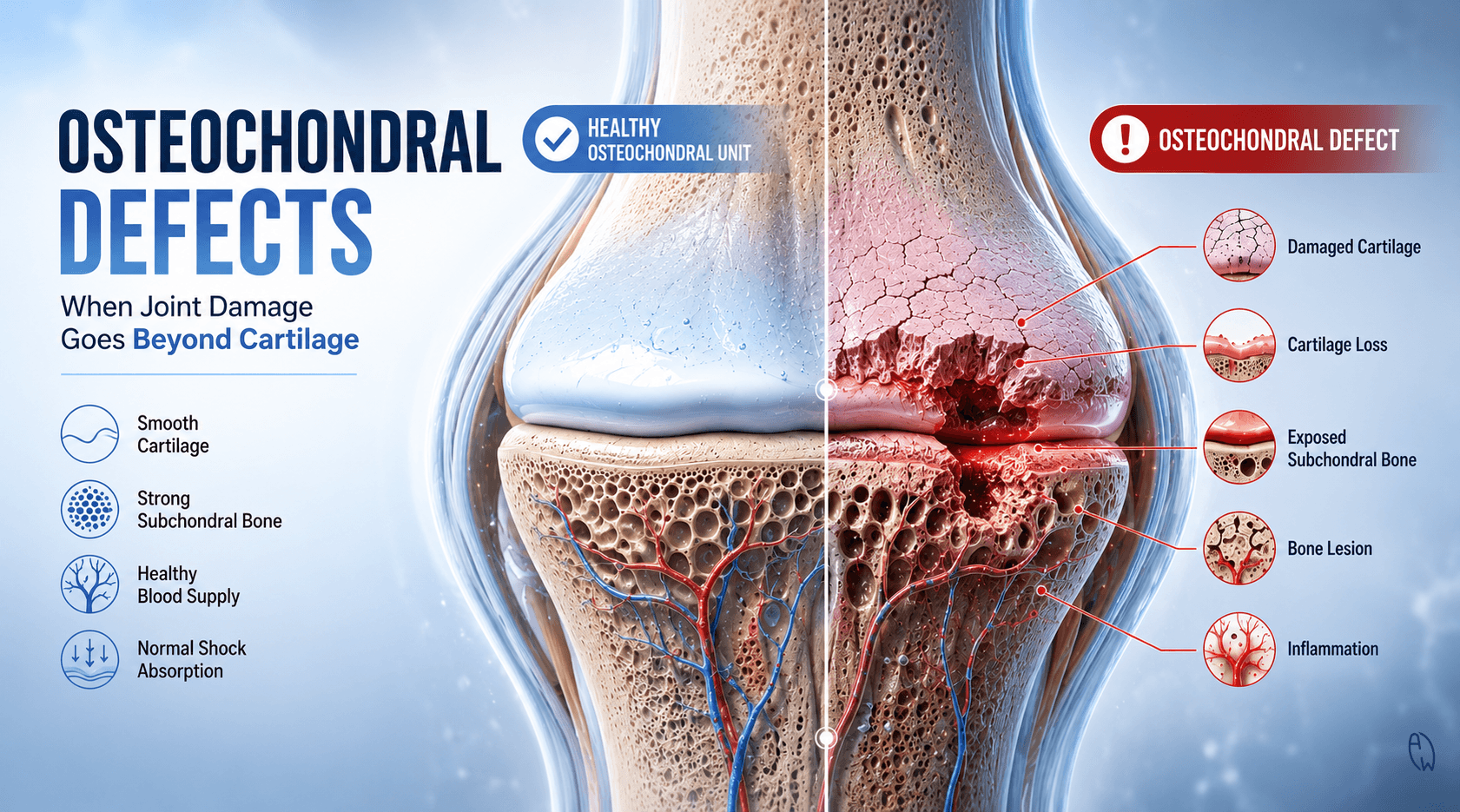
An osteochondral defect occurs when damage affects both the cartilage covering a joint surface and the underlying bone beneath it.
Because healthy movement depends on the interaction between cartilage and bone, damage to both structures can be much more challenging for the body to repair.
Whether the injury develops from trauma, repetitive stress, poor joint mechanics, metabolic health issues, or chronic degeneration, understanding osteochondral defects may help explain why some joint injuries never seem to fully heal.
What Is an Osteochondral Defect?
The word "osteo" refers to bone.
The word "chondral" refers to cartilage.
An osteochondral defect occurs when both tissues become damaged at the same location.
To understand why this matters, imagine the joint as a high-performance shock absorber.
The cartilage provides a smooth, slippery surface that allows bones to glide effortlessly against one another.
The underlying bone, known as the subchondral bone, acts as the foundation that supports the cartilage above it.
When only cartilage is damaged, the body already faces a difficult repair challenge because cartilage has very limited blood supply.
When both cartilage and bone become damaged, healing becomes even more complicated.
The result may be persistent pain, inflammation, instability, and progressive degeneration.
Understanding the Osteochondral Unit
Modern research increasingly recognizes that cartilage and bone do not function independently.
Instead, they operate as a single integrated structure known as the osteochondral unit.
This unit includes:
-
Articular cartilage
-
Calcified cartilage
-
Subchondral bone
-
Blood vessels
-
Supporting connective tissues
When one part becomes injured, the entire system may be affected.
This is why some patients continue experiencing symptoms even after the visible cartilage injury appears relatively small on imaging.
The problem often extends deeper than the surface.
Where Do Osteochondral Defects Commonly Occur?
Although osteochondral injuries can occur in many joints, they are most frequently found in areas exposed to repetitive loading and impact.
Knee
The knee is one of the most common locations.
Osteochondral defects may develop following:
-
Sports injuries
-
Twisting movements
-
Meniscus injuries
-
Ligament injuries
-
Repetitive impact loading
Because the knee experiences substantial forces during walking, running, and stair climbing, symptoms can become noticeable quickly.
Ankle
The ankle, particularly the talus bone, is another common site.
Ankle osteochondral lesions often develop after:
-
Ankle sprains
-
Repeated ankle instability
-
Sports injuries
-
Falls
Some patients continue experiencing ankle pain long after the original sprain appears to have healed.
An underlying osteochondral lesion may be the reason.
Hip
The hip joint can develop osteochondral damage from:
-
Femoroacetabular impingement (FAI)
-
Labral injuries
-
Repetitive loading
-
Degenerative changes
Shoulder and Elbow
Although less common, osteochondral injuries may also occur in athletes who repeatedly load the shoulder or elbow through throwing, lifting, or overhead activities.
What Causes Osteochondral Defects?
Many people assume these injuries occur only after major trauma.
In reality, several factors may contribute.
Acute Trauma
Direct injuries can damage both cartilage and bone simultaneously.
Examples include:
-
Falls
-
Sporting collisions
-
Sudden twisting injuries
-
High-impact accidents
Repetitive Stress
Not all damage occurs suddenly.
Years of repetitive loading may gradually weaken cartilage and subchondral bone.
This is particularly common in:
-
Athletes
-
Runners
-
Manual workers
-
Individuals with poor biomechanics
Joint Instability
When a joint moves abnormally, certain regions experience excessive loading.
Over time, these abnormal forces may contribute to osteochondral breakdown.
Degenerative Changes
As cartilage loses resilience, the underlying bone may also become compromised.
This increases the risk of developing focal osteochondral lesions.
Metabolic and Lifestyle Factors
Joint health is not determined solely by mechanics.
Nutrition, inflammation, circulation, recovery, and metabolic health also influence tissue quality.
As discussed in our article on glycation and joint aging, excess sugar consumption may contribute to chronic inflammation, collagen stiffening, and impaired tissue healing.
When Advanced Glycation End Products (AGEs) accumulate within connective tissues, cartilage may become less resilient and more vulnerable to breakdown.
You can learn more here:
How Sugar Secretly Ages Your Joints: The Hidden Damage of Glycation and AGEs
While glycation does not directly cause osteochondral defects, it may create an environment where damaged tissues struggle to recover efficiently.
Common Symptoms of Osteochondral Defects
Symptoms vary depending on the location and severity of the injury.
Deep Joint Pain
Many patients describe a deep ache located within the joint itself.
Pain often worsens during:
-
Walking
-
Running
-
Climbing stairs
-
Squatting
-
Prolonged standing
Swelling
The joint may become swollen after activity.
Swelling often improves with rest before returning again when activity resumes.
Catching or Locking
If a fragment of cartilage or bone becomes loose, patients may experience:
-
Clicking
-
Catching
-
Popping
-
Locking sensations
These symptoms should never be ignored.
Stiffness
Many individuals notice reduced range of motion and increasing stiffness, particularly after periods of inactivity.
Joint Instability
Some people report that the joint feels unreliable or weak during movement.
Why Osteochondral Defects Can Be Difficult to Heal
One reason osteochondral injuries can become chronic is the limited healing capacity of cartilage.
Unlike muscles, cartilage has very little blood supply.
This means:
-
Fewer nutrients reach damaged tissues
-
Cellular repair occurs more slowly
-
Recovery may take longer
-
Degeneration may continue despite rest
The underlying subchondral bone does receive blood flow, but healing requires healthy communication between both tissues.
If this relationship becomes disrupted, recovery becomes more difficult.
The Role of Inflammation and Recovery
Many people focus solely on the damaged joint.
However, healing depends on much more than the injury itself.
Recovery is influenced by:
-
Nutrition
-
Sleep quality
-
Circulation
-
Protein intake
-
Blood sugar control
-
Physical activity
-
Inflammation levels
A body experiencing chronic inflammation may struggle to repair cartilage and bone effectively.
This is one reason lifestyle factors can influence long-term outcomes.
Can Osteochondral Defects Lead to Arthritis?
Unfortunately, yes.
If an osteochondral lesion progresses, the smooth joint surface may become increasingly irregular.
As cartilage continues to wear, forces become less evenly distributed throughout the joint.
Over time this may accelerate:
-
Osteoarthritis
-
Cartilage loss
-
Joint degeneration
-
Reduced mobility
-
Chronic pain
This is why early recognition and proper management are important.
Supporting Joint Health and Recovery
While treatment recommendations depend on the individual case, several principles support healthier joints.
Maintain Healthy Body Weight
Reducing unnecessary loading can decrease stress on cartilage and subchondral bone.
Prioritize Protein
Protein provides the amino acids required for tissue repair and remodeling.
Control Blood Sugar
Stable blood sugar may help reduce inflammation and support tissue healing.
Stay Active
Appropriate movement helps improve circulation and joint nutrition.
Focus on Sleep and Recovery
Much of the body's repair work occurs during deep sleep.
Address Biomechanics
Joint loading patterns matter.
Poor movement mechanics can increase stress on vulnerable areas of cartilage and bone.
The Bottom Line
Osteochondral defects are more than simple cartilage injuries.
They involve a complex interaction between cartilage, bone, circulation, inflammation, biomechanics, and recovery.
Because both cartilage and the underlying bone are affected, these injuries often require greater attention than many people realize.
If you are experiencing persistent joint pain, swelling, catching, stiffness, or symptoms that never seem to fully resolve, it may be worth considering whether the problem extends beyond the surface cartilage.
Healthy joints are not maintained by movement alone.
They depend on quality nutrition, proper recovery, healthy circulation, balanced loading, and resilient connective tissues working together as one integrated system.
The stronger the foundation, the better the body's ability to withstand stress and heal when injury occurs.