Frequently Asked Questions About EOS 3D Imaging and Scoliosis
Do I need EOS 3D imaging for scoliosis?
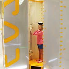
EOS 3D imaging is not mandatory for all scoliosis patients. Standard standing X-ray remains a clinically valid method for measuring Cobb angle and monitoring curves. EOS is recommended when long-term or repeated imaging is expected, especially in growing children and adolescents, to reduce cumulative radiation exposure.
Is EOS imaging safer than regular X-ray?
EOS imaging uses very low radiation, significantly less than standard X-ray and far less than CT scans. For patients who require ongoing monitoring over months or years, this reduction in radiation exposure is an important safety consideration.
Why not just use MRI instead of X-ray or EOS?
MRI is excellent for viewing soft tissues, the spinal cord, discs, and ligaments, but it is not designed to measure scoliosis curvature accurately. MRI is usually performed lying down and does not reflect how the spine behaves under gravity. For scoliosis measurement and progression tracking, standing imaging such as X-ray or EOS is required.
Why not use CT scans for scoliosis measurement?
CT scans provide detailed bone images but involve high radiation exposure and are not performed in a weight-bearing position. CT is typically reserved for surgical planning, not routine scoliosis monitoring or conservative care.
Why must scoliosis imaging be done standing?
Scoliosis is a gravity-dependent condition. Standing, weight-bearing imaging shows the spine as it functions in daily life. Supine imaging can underestimate curve magnitude and does not reflect real-world spinal loading.
Why can scoliosis measurements differ between doctors?
Differences in scoliosis measurements can occur due to:
-
Selection of different end vertebrae
-
Variations in imaging position
-
Differences in clinical purpose (surgical vs conservative care)
A variation of 3–5 degrees is recognised as normal inter-observer variability. What matters most is consistency over time, not a single number.
Why do you insist on recent X-rays or EOS images?
The Cobb angle measures the current structure of the spine. Growth, posture, degeneration, and compensation can change curvature over time. Using outdated images risks inaccurate planning. Recent imaging ensures we are measuring the spine as it exists now, not months or years ago.
Is EOS imaging worth the higher cost?
EOS imaging is typically around three times more expensive than standard X-ray. If budget is a concern, standard X-ray is an acceptable alternative. When cost is not a limiting factor and repeated imaging is expected, EOS offers the advantage of very low radiation while maintaining measurement accuracy.
Will imaging guarantee scoliosis improvement?
No imaging method guarantees outcomes. Imaging guides measurement, monitoring, and clinical decision-making, but the body’s response varies between individuals. We do not promise results beyond what the body can safely achieve.
Can you treat everything seen on imaging?
No. If imaging reveals conditions outside conservative scoliosis care — such as acute injury, neurological involvement, or ligament damage — we will refer you to the appropriate medical specialist. Ethical care includes knowing when not to treat.
Imaging does not treat scoliosis — accurate measurement guides safe care.
That is why we prioritise standing imaging, consistent Cobb angle measurement, and the lowest reasonable radiation exposure over time.