EOS Imaging for Scoliosis: Elevating Diagnosis & Treatment
Scoliosis is more than a simple sideways curve of the spine. It is a three-dimensional spinal condition involving lateral curvature, vertebral rotation, and changes in overall body balance. Accurate imaging is essential to diagnose scoliosis, monitor curve progression, and guide safe treatment decisions—especially in children and adolescents who are still growing.
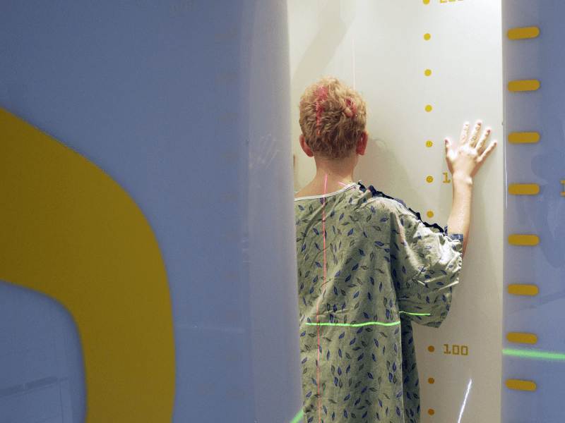
One of the modern imaging technologies increasingly used in spinal assessment is the EOS Imaging System. This advanced system allows clinicians to evaluate the spine in a standing, weight-bearing position while using significantly lower radiation exposure compared with conventional imaging.
While EOS technology offers important advantages, it is also important to understand that standard standing X-rays remain a reliable and clinically accepted method for measuring scoliosis curves. The choice of imaging depends on the patient's clinical needs, monitoring frequency, and overall treatment plan.
The Evolution of EOS Imaging
The development of the EOS Imaging System has significantly advanced orthopedic imaging and the way clinicians evaluate skeletal alignment and spinal deformities.
EOS technology was developed to address a key challenge in musculoskeletal medicine: providing accurate imaging while minimising radiation exposure, especially for children who may require repeated scans during growth.
Research published in the European Journal of Orthopaedic Surgery & Traumatology highlights the innovative design of this imaging technique. EOS uses a simultaneous biplanar radiographic scanning system, meaning it captures two images at the same time:
-
A frontal view (anterior–posterior)
-
A side view (lateral)
Capturing these two images simultaneously allows clinicians to evaluate skeletal alignment efficiently while reducing radiation exposure compared with traditional imaging approaches.
Another major advantage of EOS imaging is its ability to generate full-body standing images. This gives doctors a comprehensive view of the entire musculoskeletal system, allowing them to evaluate how different parts of the body interact, including:
-
Spine alignment
-
Pelvic balance
-
Hip positioning
-
Knee alignment
-
Leg length relationships
This broader perspective is particularly valuable when managing complex conditions such as scoliosis, where the spine, pelvis, and lower limbs often influence each other.
EOS Referrals and Exams
EOS imaging is primarily used to assess patients with spinal, hip, and lower limb alignment disorders.
It is particularly beneficial for children and adolescents with progressive spinal conditions, including scoliosis and other spinal deformities that require frequent monitoring over time.
However, EOS imaging is not typically used for routine injuries that can be evaluated using standard radiography. For example:
-
Broken bones in the arms
-
Fractures in the legs
-
Injuries involving the hands or feet
For these conditions, traditional X-rays remain the standard diagnostic method.
How EOS imaging referrals work
For patients who require EOS imaging as part of scoliosis evaluation or monitoring, referrals may be made to Posture Ray Imaging Centre, where low-dose standing 3D musculoskeletal imaging is available.
Why Imaging Is Necessary in Scoliosis
Scoliosis is a three-dimensional deformity of the spine, involving curvature, vertebral rotation, and changes in body alignment.
Doctors monitor scoliosis using the Cobb Angle, which measures the severity of spinal curvature and helps determine whether the condition is progressing.
Imaging is necessary to:
-
Diagnose scoliosis
-
Measure the Cobb angle
-
Monitor curve progression during growth
-
Evaluate treatment outcomes
-
Guide clinical decision-making
Because scoliosis patients may require multiple scans over several years, the type of imaging used becomes an important consideration.
Why Radiation Exposure Matters in Scoliosis
Children with scoliosis often require repeated imaging during growth, sometimes over several years.
Although standard X-rays use relatively low radiation, repeated exposure over time is still a clinical consideration.
The EOS Imaging System was developed to reduce this cumulative exposure while maintaining accurate spinal measurement.
For this reason, EOS imaging is often considered beneficial for young patients who require long-term scoliosis monitoring.
Why Scoliosis Imaging Must Be Done Standing
Scoliosis is a gravity-dependent condition.
The curvature of the spine changes depending on whether the body is lying down or standing under normal body weight.
Standing imaging allows clinicians to observe how the spine behaves during daily activities such as standing and walking. Imaging performed in a lying position can underestimate the true curve magnitude.
Both EOS imaging and standard scoliosis X-rays are performed in a standing position to capture the spine under normal weight-bearing conditions.
Frequently Asked Questions About EOS Imaging
Do I need EOS imaging for scoliosis?
EOS imaging is not required for all scoliosis patients. Standard standing X-rays remain a reliable and clinically accepted method for measuring spinal curvature.
EOS imaging is often recommended when long-term monitoring is expected, particularly in growing children who may require repeated imaging.
Is EOS imaging safer than standard X-rays?
EOS imaging uses significantly lower radiation doses than conventional radiography and much less than CT scans. This reduced exposure can be beneficial for patients who need frequent imaging over time.
Why not use MRI instead?
MRI is excellent for evaluating soft tissues such as the spinal cord, discs, and ligaments, but it is usually performed while the patient is lying down. Because scoliosis is influenced by gravity, MRI is not the best tool for measuring spinal curvature progression.
Why not use CT scans?
CT scans produce detailed bone images but involve much higher radiation exposure and are not performed in a weight-bearing position. They are generally reserved for surgical planning or complex spinal conditions.
Why can scoliosis measurements vary between doctors?
Small differences in Cobb angle measurements may occur due to:
-
Selection of different end vertebrae
-
Variations in patient positioning
-
Differences in measurement technique
A difference of 3–5 degrees is considered normal clinical variation. Consistent monitoring over time is more important than focusing on a single measurement.
The Role of Imaging in Responsible Scoliosis Care
Imaging does not treat scoliosis — accurate measurement guides safe care.
Whether using standard standing X-rays or the EOS Imaging System, the goal of scoliosis monitoring is to balance three important priorities:
-
Accurate Cobb angle measurement
-
Consistent monitoring of spinal changes
-
The lowest reasonable radiation exposure
When used appropriately, modern imaging technologies help clinicians understand spinal structure more clearly and design safe, personalised treatment strategies for scoliosis patients.
References
Medical Disclaimer
This article is for educational purposes only and does not constitute medical advice.
Scoliosis varies significantly between individuals. Always consult a qualified healthcare professional before starting any new sport or exercise program, especially if you have scoliosis, spinal conditions, pain, or previous injuries. Participation in sports should be guided by individual assessment and professional recommendation.
The image is shared for educational purposes with patient consent. Individual outcomes vary. Structural correction does not automatically restore full respiratory function. Clinical assessment is required.
Copyright & Content Protection Notice
© 2026 All Well Scoliosis Centre. All rights reserved.
This content is original and protected by copyright law.
No part of this article may be reproduced, distributed, copied, or reused in any form without prior written permission. Unauthorized use, duplication, or content scraping is strictly prohibited.